

Discover more about Menarini Group
All company structures are involved at different levels in the drug manufacturing process...
MARKETS
CENTRAL AND EASTERN EUROPE

information reserved for healthcare professionals
The information contained in this section is intended for the exclusive use of healthcare professionals.
Zenit Fast
The information to be found in this section is intended for the exclusive use of healthcare professionals.
- FEATURES
- TECHNICAL DATA
- LEARN MORE
- MULTIMEDIA

As fast and simple as you want it to be
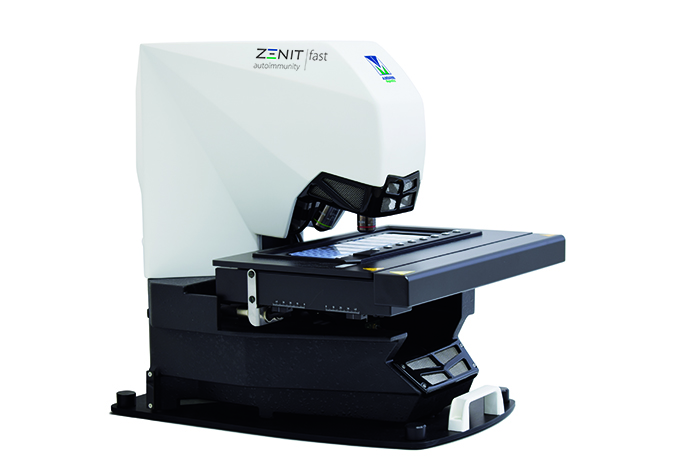
Zenit fast is the new A. Menarini Diagnostics automated system for the acquisition, analysis and storage of IFA slides, which provides laboratories with an efficient solution to improve the management of IFA tests, standardize the validation procedure and reduce intra- and inter-laboratory variability.
What distinguishes Zenit fast is the image autofocusing technology, whole well scanning, the software algorithms for IFA detection and pattern recognition, run-time, types of ANA IFA patterns recognized and its ability to analyze different IFA substrates.
Main features and benefits
- Fast and efficient screening of negative and positive samples
- Fast slide scanning: <30 seconds per well (HEp-2 slides, 20x), >2.5 mins per well for tissues
- Multirun sessions to process large sample numbers
- 8-slide tray
- High-quality scanning sensor and LED microscope illumination
- Whole well scanning
- Virtual microscope for well navigation
- Reduced operating costs
Powerful and reliable information management
Zenit fast offers powerful analytics with a simple and intuitive software interface.
The software is structured into 3 main modules: Scanning, Validation, and Archive.
- Scanning: The user can upload a worklist and launch the automatic scanning of slides that are already mounted and ready on the microscope stage. Scanning can be launched in manual or automatic mode.
- Validation: The digitized image is displayed and can be navigated with the virtual microscope tool. Results are interpreted and validated by the operator using the software interface.
Customizable views facilitate navigation. Results can be sent directly to LIS as soon as each well is validated. - Archive: The user can access the patient database, performed tests and processed slides. Free text search or multiple and complex queries can be set.
Automated image analysis software
The system performs automatic classification of positive/negative results for Zenit ANA HEp-2 tests measuring the intensity of fluorescence for each positive test and provides pattern recognition on positive results (see detected patterns in the Technical Data section). Mitosis recognition is also performed on identified patterns.
The system is now also capable of discriminating between positive and negative samples for both ANCA E and ANCA F and to define the type of pattern (see detected patterns in the Technical Data section).
A new feature in the software allows also the discrimination between positive/negative results on DNA Crithidia slides.
Magnification
4x, 20x
Optional magnification
Three extra positions to introduce standard microscope objectives, up to five loaded objectives. Maximum magnification 40x
SCANNING SPEED
Less than 30 seconds/well for ANA HEp-2 slides at 20x
Less than 2.5 minutes/well for tissue slides at 20x
Resolution at 20x
0.5 micron
Scanning mode
Single or multiple runs per session
Type of compression
Standard JPEG-2000
Compression level
ANA/ANCA/nDNA: 5-10 MB per well; tissues: 100-150 MB per well
2D Data matrix reader
Yes
Connectivity
Bidirectional, HL7 protocol
Scanning capability
HEp-2, ANCA, nDNA, tissues
Automated analysis
Zenit ANA HEp-2 pattern recognition (8 patterns identified): AC-0 Negative, AC-1 Nuclear homogeneous, AC-2,4,5,29 Speckled, AC-3 Centromere, AC-6,7 Nuclear dots, AC-8,9,10 Nucleolar, AC-15,16,17,18,19,20,22,23 Cytoplasmic, AC-21 Cytoplasmic reticular AMA
Dedicated gallery of mitoses on recognized patterns and relocalization
Zenit ANCA ethanol and ANCA formalin pattern recognition (Negative, c-ANCA, p-ANCA, Others).
Zenit nDNA positive/negative recognition.
Atlas / Pattern guide
Reference images with ANA Nomenclature ICAP codes
Electrical specifications
Power source: AC 100-240V 50/60 Hz
Power consumption: 45 VA
Mechanical specifications
Scanner: Height: 50 cm / Width: 60 cm / Length: 40 cm / Weight: 30 kg
Processing unit: Height: 46 cm / Width: 52 cm / Length: 21 cm / Weight: 15 kg
Display unit: Inches: 24” / Resolution: 1920x1200 / Height: 46-55 cm / Width: 25 cm / Length: 57 cm / Weight: 10 kg
All Zenit slides are compatible with the A. Menarini Diagnostics Zenit series platforms and can be assessed either visually with a fluorescence microscope or automatically with the A. Menarini Diagnostics interpretation system Zenit fast, Zenit PRO or Zenit G-Sight.
CHARACTERISTICS
- Significant number of mitotic cells
- Optimized cell morphology
- Large nuclei for easy pattern recognition
- Optimal substrate for the detection of antibodies targeting cytoplasmic antigens
- ANA positive controls (different patterns) provided
- Substrate provided in accordance with CLSI standard guidelines
ORDERING INFORMATION
Our products are conveniently available in all parts of Europe through the network of local A. Menarini Diagnostics Subsidiaries. Please contact your nearest one for additional information and order placing.
ICAP Nomenclature for ANA Patterns
The nomenclature and classification tree for all HEp-2 cell patterns as shown at www.ANApatterns.org is shown here below.
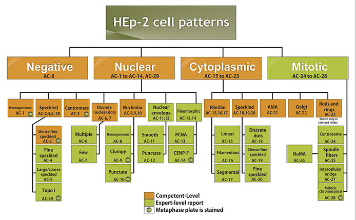
Chan, E. K. L., J. Damoiseaux, O. G. Carballo, K. Conrad, W. de Melo Cruvinel, P. L. Francescantonio, M. J. Fritzler, I. Garcia-De La Torre, M. Herold, T. Mimori, M. Satoh, C. A. von Muhlen and L. E. Andrade (2015). "Report of the First International Consensus on Standardized Nomenclature of Antinuclear Antibody HEp-2 Cell Patterns 2014-2015." Front Immunol 6: 412.
There are total of 30 ICAP patterns designated with alphanumeric AC code for each from AC-0 to AC-29. Boxes with amber background are recommended as competent-level reporting, whereas those with olive green background are considered for expert-level reporting. AC stands for anti-cell.
A.Menarini Diagnostics Autoimmunity platforms (including Zenit fast) follow ICAP nomenclature and classification
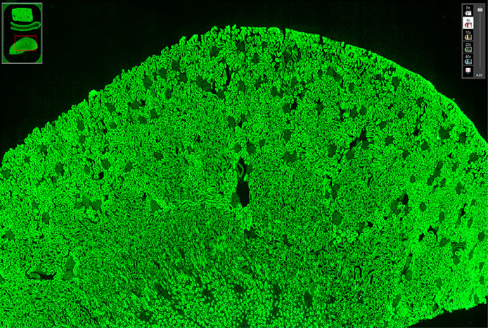
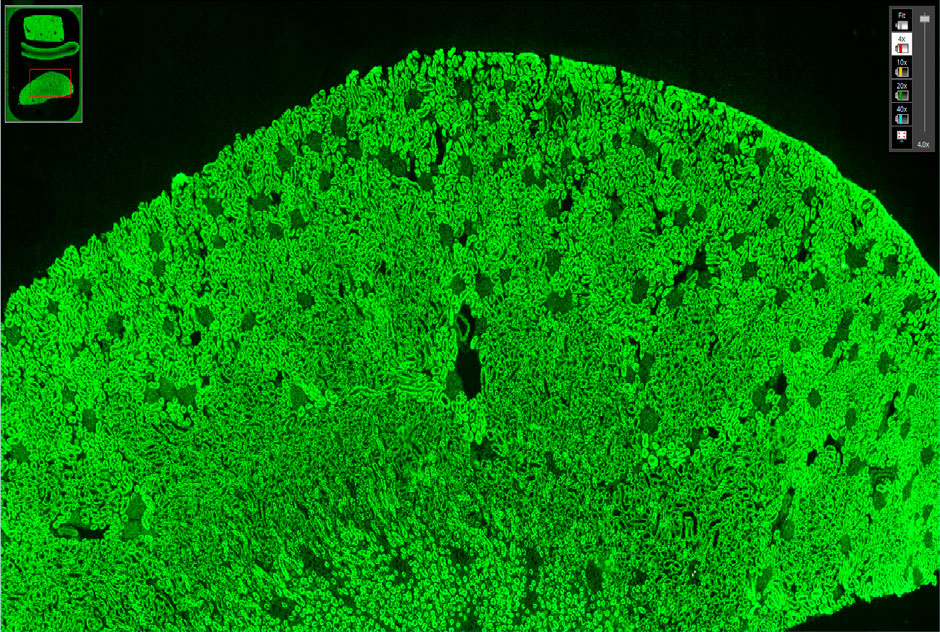 Full screen view of the IFA test: the image can be navigated with the virtual microscope.
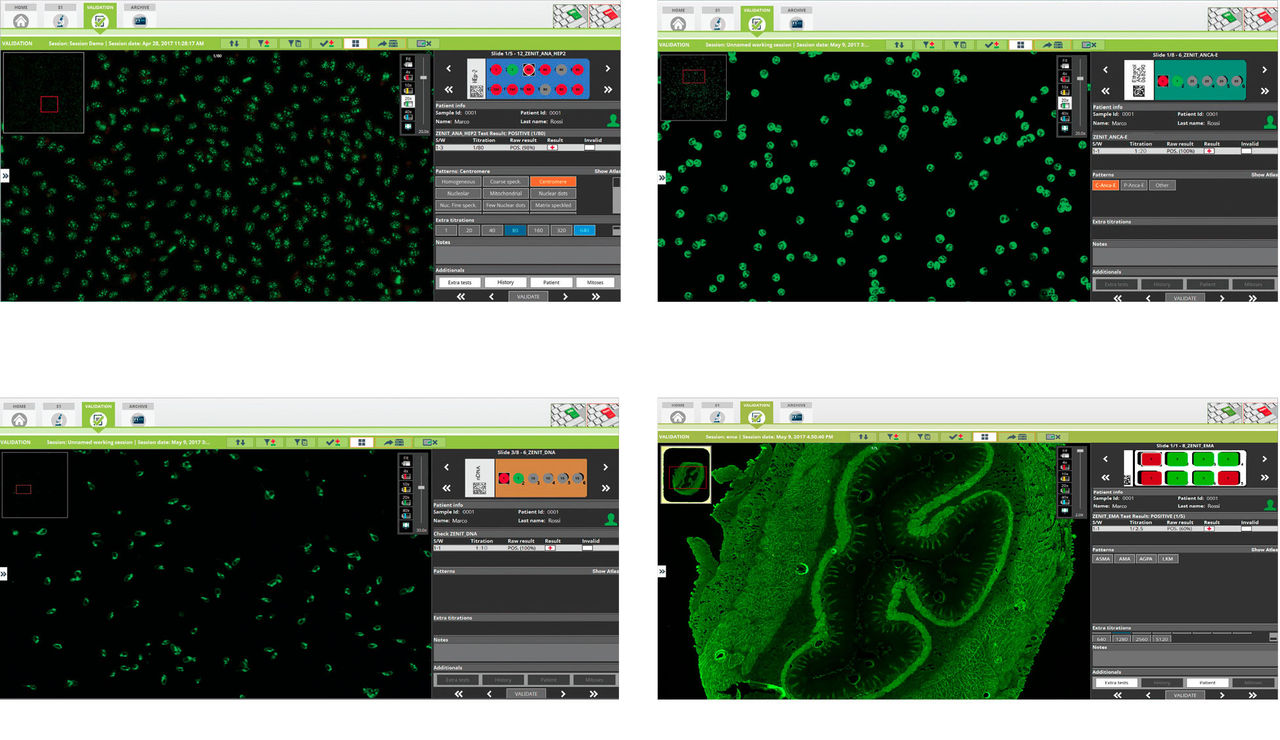
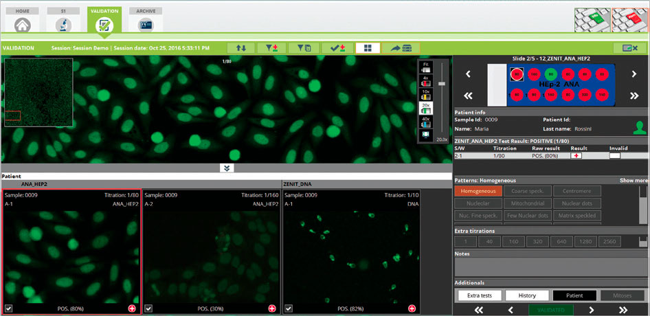 Multiview in the Validation module with multiple wells of the same patient’s sample.
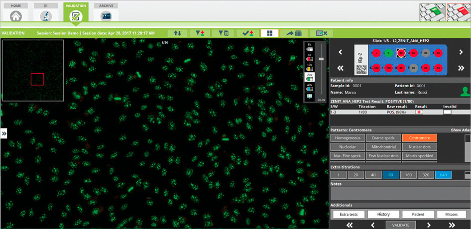 Zenit ANA HEp-2 pattern recognition
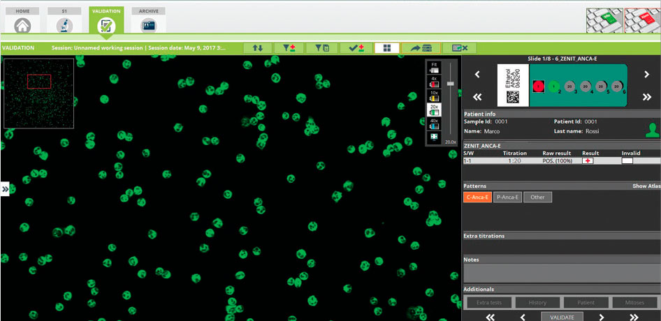 Zenit ANCA positive/negative classification and pattern recognition
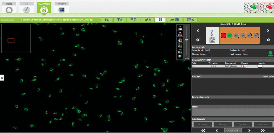 Zenit nDNA positive/negative classification
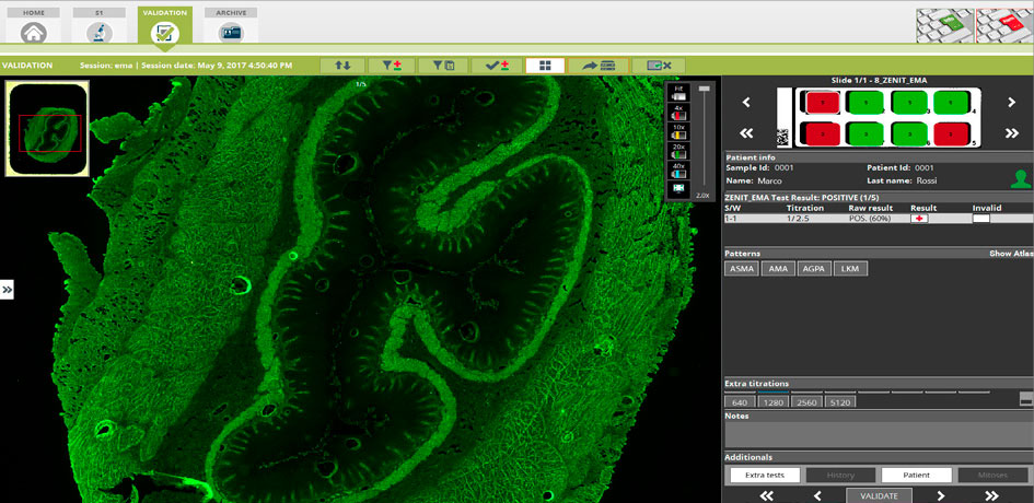 Zenit EMA positive/negative classification