

Discover more about Menarini Group
All company structures are involved at different levels in the drug manufacturing process...
MARKETS
CENTRAL AND EASTERN EUROPE

information reserved for healthcare professionals
The information contained in this section is intended for the exclusive use of healthcare professionals.
Zenit G-Sight
WHAT IS ZENIT G-SIGHT
WHAT IS ZENIT G-SIGHT
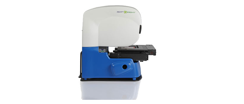
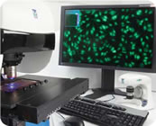
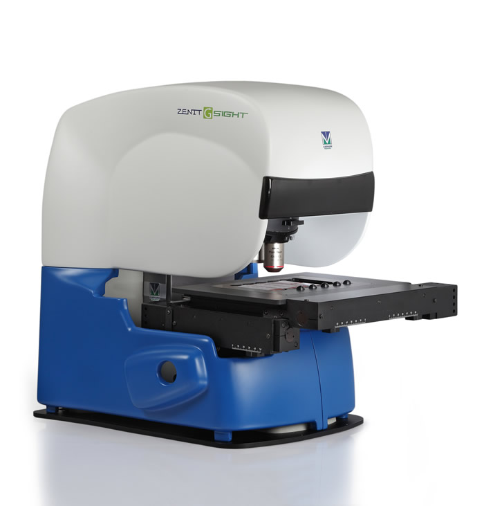
Zenit G-Sight is the novel A. Menarini Diagnostics automated system for image acquisition and interpretation of cell- or tissue-based indirect immunofluorescence (IIF) tests.
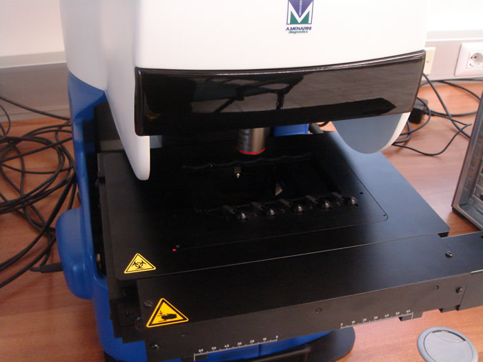
Components of this compact benchtop instrument include:
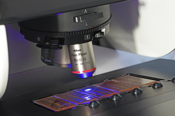
- a state-of-the-art automated fluorescence microscope with LED light source
- a motorized scanning stage for substrate slides
- a highly sensitive CCD camera
- an innovative software for digital image generation and analysis
Zenit G Sight performs:
- Fully automated image acquisition of cell- or tissue-based IIF tests
- Semi-quantitative analysis and intelligent pattern recognition for ANA HEp-2 assays
- Digitization for other IIF substrates
- Analysis and intelligent pattern recognition for other IIF substrates available soon
Clarification of suspected rheumatic symptoms is mainly achieved by the serological assessment/detection of disease-specific autoantibodies (e.g., anti-nuclear antibodies, ANAs).
Despite the development of enzyme-linked immunosorbent immunoassay (ELISA) and other alternative methods or technologies, screening for ANAs by IIF assays remains the gold standard method in the current diagnostic approach to systemic rheumatic disorders such as systemic lupus erythematosus (SLE), Sjögren’s syndrome (SS), progressive systemic sclerosis (PSS), dermatomyositis-polymyositis (DM/PM) or mixed connective-tissue disease (MCTD).
International guidelines recommend that ANA screening be performed on human epithelial (HEp-2) cells. Other cell substrates such as Crithidia luciliae (for the detection of autoantibodies to dsDNA) and human granulocytes (for the detection of anti-neutrophil cytoplasmic autoantibodies, ANCA) are routinely employed in cell-based IIF procedures.
Since autoantibodies produce characteristic fluorescence patterns whose analysis depends on the knowledgeability and the subjective interpretations of investigators, high intra- and interlaboratory variability represents a major problem in autoimmune diseases diagnostics.
The increase in workload and the need for standardized and reproducible evaluation of cell-based IIF assays have
guided autoimmunity laboratories on the road to automation.
The new Zenit G Sight system from A. Menarini Diagnostics was specifically designed to overcome IIF-specific issues, making it particularly suited for the automated serological diagnosis of autoimmune diseases in a routine laboratory setting.
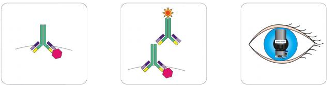
IIF ASSAY TPROCEDURE
The principal steps of the method are:
- Serum sample dilution (for example 1:80) and incubation with substrate fixed on glass slides for 30 min in a moisture chamber at RT
- Incubation with fluorescein isothiocyanate (FITC)-conjugated sheep anti human Ig for 30 min at RT
- Assessment: visual or automated analysis of slides
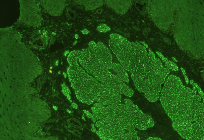
IIF PATTERNS*
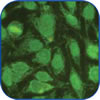
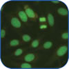
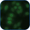
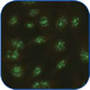
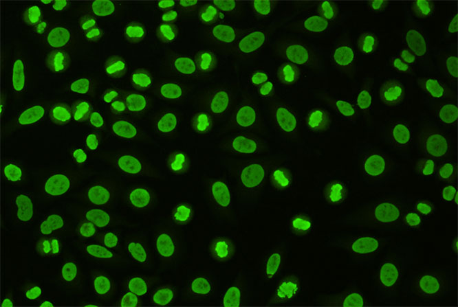
ANA detection on HEp-2 cells generates specific fluorescence patterns. The five main patterns which can be found are the following
 Homogeneous/Peripheral
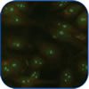 Nucleolar
 Speckled
 Centromere
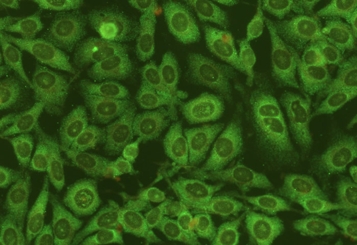
 Cytoplasmic/ Mitochondrial
* ANA fluorescence patterns on HEp-2 cells are shown because this is the most widely used test for visual/automated screening of rheumatic diseases
IIF assay is a low-cost, simple and rapid method for routine autoimmune screening; however, it has several disadvantages, which can be easily resolved with Zenit G Sight.
Classical cell-based IIF assay
- Subjective analysis
- Limited number of samples
- Time-consuming sample screening
- Fluorochrome decay
- Organized archiving space required
- Difficult to exchange results with other labs
vs Zenit G Sight system
- Objective quantification and pattern recognition
- High workload management
- Efficient automated +/- sera discrimination
- Digitized image data
- File storage and easy retrieval of previous reports
- Online access and data exchange
ZENIT G-SIGHT IS:
- Valuable and accurate
giving clinicians a precious aid for diagnosis by providing a full quantitative/qualitative report on autoantibody serological status
- Reliable
providing results that are reproducible and comparable within and between laboratories
- Cost-effective
alleviating the heavy workload of laboratories with large sample numbers, maximizing efficiency and optimizing human resources
- Secure
safeguarding results and improving data management
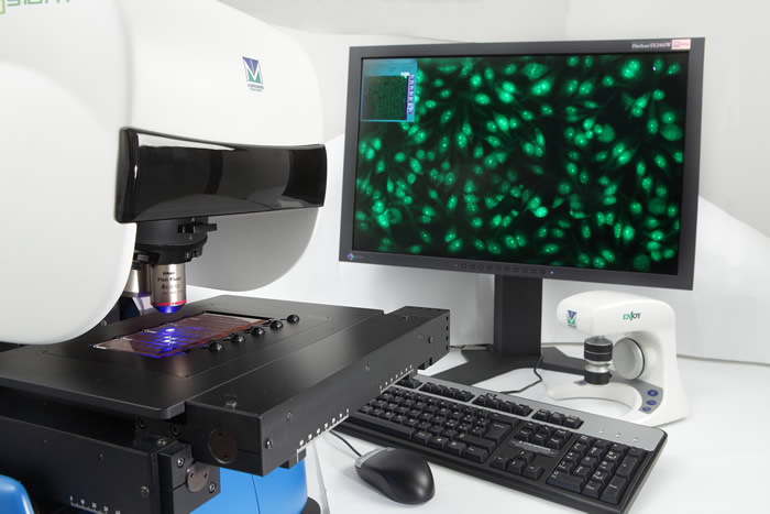
The innovative Zenit G Sight software is the central component of the system. It guarantees optimal image capture and processing, fully automated analysis of immunofluorescence signals and efficient data storage and management. In addition, Zenit G Sight software manages and controls the entire experimental workflow.
DIGITAL IMAGE ACQUISITION
Zenit G Sight software automatically reads out images and generates editable high-resolution Jpeg2000 files (30-50 MB per well). Enlarged images can subsequently be obtained by means of digital magnification.
IMAGE INTERPRETATION / EVALUATION
Zenit G Sight software processes the fluorescence signals and ensures an objective reading: it automatically analyzes signal intensity and distribution in each sample.
A cut-off level was set in order to minimize the risk of “false negatives”. After the positive-negative differentiation step, a mathematical algorithm automatically assigns the observed fluorescence, for example, to one of the five basic patterns in the case of HEp-2 substrate.
The system can also give a semi-quantitative result on a positive identification criteria basis (for the HEp-2 cells, the dsDNA-mAb33 Cat. Number 33300 vial 10000 IU correlated with WHO standard Wo/80 has been used as reference material, to estimate the Absolute concentration/fluorescent emission rate).
SOFTWARE
Image storage
Once the manual validation process is completed, Zenit G Sight software proceeds with immunofluorescence image archiving. The system provides sufficient data storage capacity for about 5000 slides*.
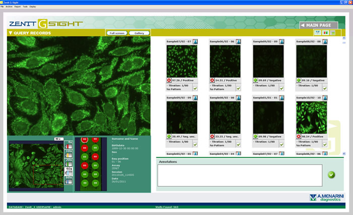
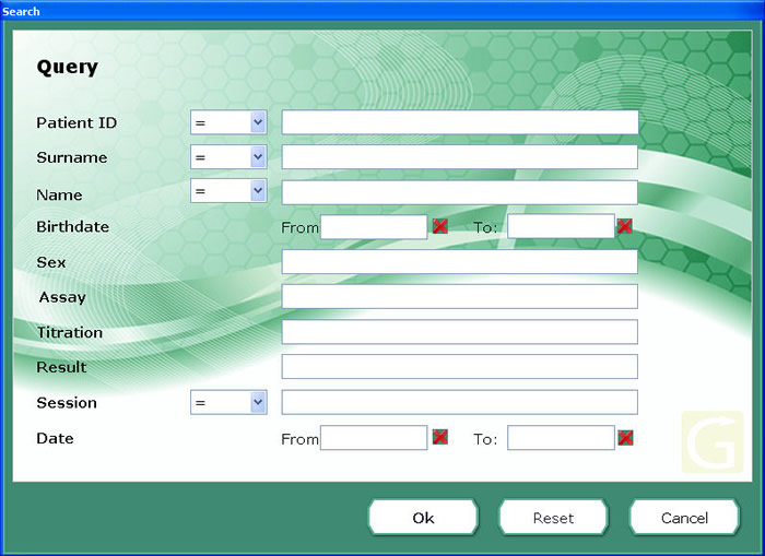
Data retrieval and sharing
The Zenit G Sight software database search option allows easy retrieval of previous results.
Online data access and exchange are also possible.
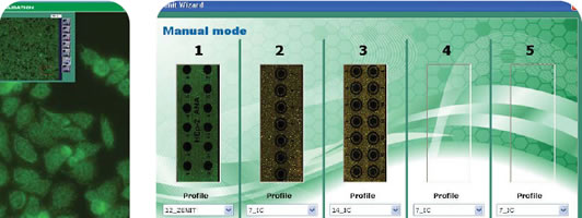
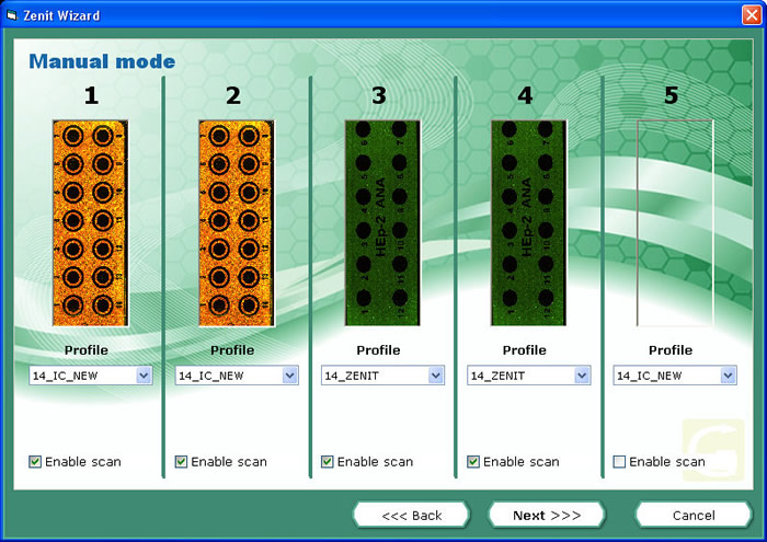
Workflow management
The intuitive, user-friendly interface of Zenit G Sight software accompanies the operator step by step through the entire autoantibody detection process:
1. Worksheet loading (automatic or manual)**
The worksheet contains the number of slides to process, the test to be carried out for each slide and sample data (for each well: patient identification number, patient details, antibody title etc…).
2. Scanning and automatic analysis of samples
After slides are placed on the stage, the system automatically acquires images and proceeds to analyze them using a specific algorithm to discriminate between positive and negative samples and assign a pattern to each well.
3. Validation
The operator examine every digital image and navigate the virtual slide. Moreover the diagnosis provided by the system can be confirmed or modified based on the clinical and laboratory history of the patient.
4. Report (to print or as an export file)
The system allows report printing and creation of an output worksheet file containing the pattern and associated diagnosis for every well.
* depends on the HDD capacity;
** Zenit G Sight can be connected to LIS and to all Zenit series A. Menarini Diagnostics liquid handling systems for the automatic preparation of IIF slides
Technical specifications
Objectives
4x, 40x
Optional objectives
Four remaining available positions for accommodating objectives (max. of 6)
Scanning speed
15-60 minutes for a 14 wells slide
Optical source
LED
Type of compression
Standard JPEG-2000
Compression level
30-50 MB per well
Environmental setting
Work session
Temperature
0-40 °C
Humidity
85% RH max. (without condensation)
Transport/storage
Temperature
from -20 to 60 °C
Humidity
90% HR máx. (without condensation)
Power supply
Entry level
AC 110-120 V or 220-240V 50-50 Hz
Physical characteristics
Zenit G-Sight (Microscope)
Dimensions: L 40 cm x H 60 cm x W 60 cm
Weight: 40 kg
Elaboration unit (PC)
Operating system: Windows XP SP3 Professional
Processor: Intel Xeon QCore
Live memory: 4GB DDR3
Hard Disk: 4x500 GB SATA RAID10 STRIPED/MIRRORED
Dimensions: L 21 cm x H 46 cm x W 52 cm
Weight: 20 kg
Visual display unit (Monitor)
Monitor: EIZO LCD
Screen size: 24”
Screen resolution: 1920 x 1200 pixels (wide screen format)
Dimensions: L 57 cm x H 46-55 cm x W 25 cm
Weight: 12 kg
LISTING
Zenit G-Sight: analyzer code 42251
Zenit G-Sight: Enjoy code 42677
Zenit G-Sight: full viewer license code 42679
Zenit G-Sight: additional working station MAC code 42676
Zenit G-Sight: OBJ.20x code 42678
ZENIT G-SIGHT AT A GLANCE
- Compact benchtop system
- Fully automated image acquisition of cell- or tissue-based indirect immunofluorescence (IIF) tests
- Semi-quantitative analysis and intelligent pattern recognition for ANA HEp-2 assays
- Digitization for other IIF substrates
- Analysis and intelligent pattern recognition for other IIF substrates available soon
- High workload management
- Data storage and retrieval
- Online remote access and file sharing
TEST AVAILABLE
HEp-2 CELL SUBSTRATE SLIDES
All our substrate slides locally distributed are compatible with the A. Menarini Diagnostics Zenit series platforms and can be assessed either visually with a fluorescence microscope or automatically with the A. Menarini Diagnostics interpretation system Zenit G Sight.
CHARACTERISTICS
- Significant number of cells in mitosis
- Optimized cell morphology
- Large nucleus for easy pattern recognition
- Optimal substrate for the detection of antibodies targeting cytoplasmic antigens such as antigens associated with ribosomal P, tRNA synthetase (Jo-1), mitochondria and smooth muscle
- ANA positive control provided
- Substrate provided in accordance with CLSI standard guidelines
ORDERING INFORMATION
Our products are conveniently available in all parts of Europe through a network of local A. Menarini Diagnostics branches. Please contact your nearest branch for additional information and order placing
DIAGNOSTIC SIGNIFIANCE OF ANTI-NUCLEAR ANTIBODIES
IIF staining pattern
Nature of antigen
Associated disease
Homogeneous/
Peripheral
Deoxyribonucleoprotein DNA
SLE with renal involvement SLE

Nucleolar
4S-6S RNA probably U3
RNA
Scleroderma
Speckled
RNP
Sm
SS-A/SS-B
Scl-70
SLE or Mixed Connective Tissue Disease
SLE
SLE or Sjögren’s Scleroderma

Centromere
Inner and outer plates of kinetochore
CREST Syndrome
Cytoplasmic/
Mitochondrial
70 kd mitochondrial protein
primary biliary cirrhosis
Anti-nuclear antibody (ANA) detection: Zenit G Sight vs visual assessment
ANAs were determined by IIF using HEp-2 cells as a substrate. Tests were conducted blinded on 120 sera from patients with Connective Tissue Diseases or other autoimmune diseases and healthy subjects. Results were evaluated by three independent expert physician examiners and compared with evaluation by the Zenit G Sight automated interpretation system. Excellent agreement (100%) was obtained for the differentiation of positive and negative samples. Discrepancies were observed only in sera identified by Zenit G Sight system as “doubtful negative”: 8% of these were reported “positive speckled” by examiners.
- ANCA slides
PHOTO GALLERY
For additional information, please visit the following web site: http://gsight.eu/
Please be informed that the links above will take you to a website the content of which has been designed in accordance with country specific laws and regulations.